Corneal Topography: Everything You Need to Know About Corneal Mapping
Published: July 6, 2025
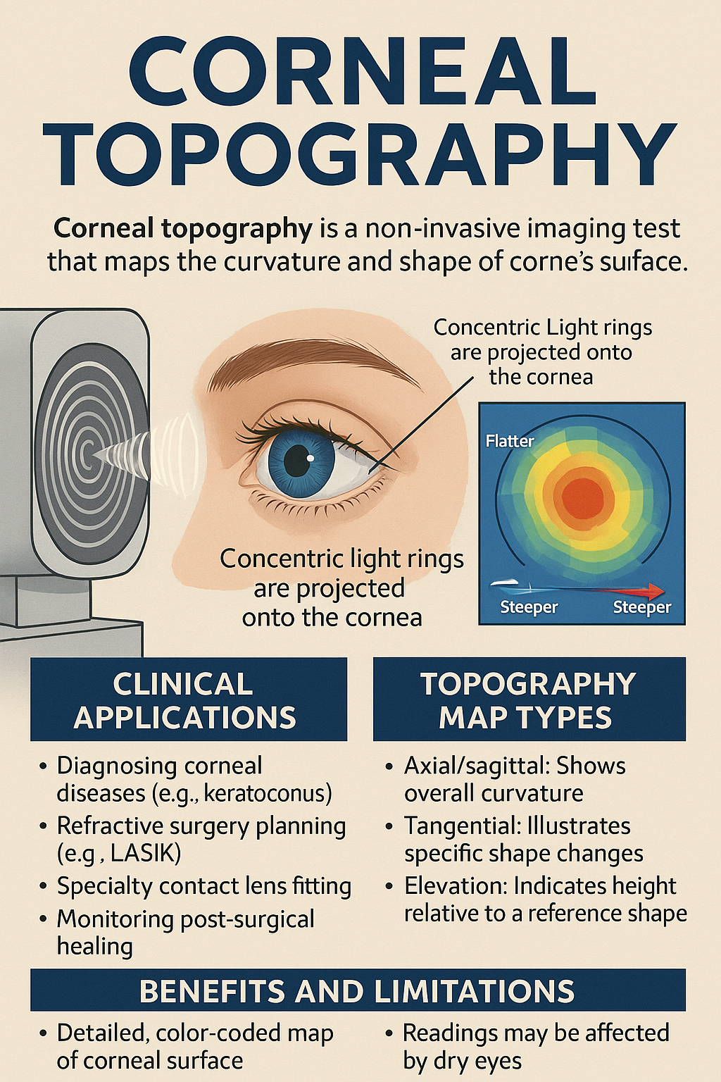
Corneal topography is a cutting-edge, non-invasive imaging test that creates a detailed, color-coded map of the cornea’s surface curvature and elevation. It’s invaluable for diagnosing corneal disorders, planning vision correction surgery, fitting specialty contact lenses, and monitoring postoperative healing. This guide covers how corneal topography works, why it’s performed, what the results mean, and what to expect as a patient.
How Corneal Topography Works
At its core, corneal topography measures the shape of the anterior corneal surface. Different technologies are used:
- Placido-ring systems project concentric light rings onto the cornea and record their reflections to calculate curvature.
- Scheimpflug imaging uses a rotating camera and slit beam to capture cross-sectional “slices,” measuring both curvature and elevation—even when the cornea is cloudy.
- Swept-source OCT topographers employ high-speed light sources to generate ultra-high-resolution 3D maps of anterior and posterior surfaces.
Clinical Applications
Corneal topography has revolutionized anterior segment care. Key uses include:
- Keratoconus screening & monitoring: Early detection of cone-shaped protrusions and thinning; tracking progression to determine timing for treatments like corneal cross-linking.
- Refractive surgery planning: Safe LASIK/SMILE/PRK candidacy assessment by measuring corneal thickness (pachymetry) and detecting irregular astigmatism.
- Specialty contact lens fitting: Designing rigid gas-permeable, scleral, or orthokeratology lenses that match complex corneal profiles for comfort and optimal vision.
- Postoperative follow-up: Monitoring corneal healing after surgery or cross-linking, detecting early ectasia or aberrations.
Interpreting Your Topography Maps
Maps are displayed in color gradients: warm hues (red/orange) mark steeper or elevated areas, cool hues (blue/green) flatter regions. Common map types:
- Axial (Sagittal) Curvature: Shows average curvature radiating from the corneal apex—good for general screening.
- Tangential (Instantaneous) Curvature: Highlights local curvature changes—ideal for pinpointing early keratoconus.
- Elevation Maps: Compare corneal surface to a best-fit sphere or toric reference—detect subtle bulges or depressions.
- Pachymetry Thickness Map: Overlay of corneal thickness—critical for refractive surgery safety and keratoconus staging.
What to Expect During Your Exam
The test is quick (1–3 minutes), painless, and involves no eye contact:
- You rest your chin on a support and look at a fixation target (a red or green light).
- The device projects rings or light patterns and captures images.
- No drops are required unless combined with other tests like pachymetry.
- The examiner reviews multiple maps for consistency—sometimes repeating scans for clarity.
Benefits and Limitations
Benefits: Non-contact, fast, highly detailed data; critical for early disease detection and personalized treatment. Limitations: Tear film instability (dry eye) can distort readings; severe corneal scarring may reduce accuracy. Combining topography with tomography or OCT ensures comprehensive evaluation.
Preparing for Your Appointment
To ensure accurate results:
- Avoid wearing soft contact lenses for 24–48 hours or rigid lenses for up to a week before your exam.
- Keep your eyes well-lubricated; use preservative-free artificial tears if needed.
- Remove eye make-up and avoid creams or lotions that could smear on the skin.
Future Trends in Corneal Imaging
Emerging advances include:
- AI-driven analysis: Automated detection of keratoconus and risk prediction software.
- Combined multi-modal platforms: Integration of topography, tomography, and biomechanical metrics for comprehensive corneal profiling.
- Portable devices: Hand-held scanners for screening in remote or underserved areas.
Conclusion
Corneal topography has become an indispensable part of modern eye care, offering unparalleled insight into corneal health. Whether you’re evaluating for surgery, managing keratoconus, or fitting specialty lenses, this detailed mapping ensures personalized, precise treatment. Talk to your eye care professional to see if corneal topography is right for you.
Keyphrase: corneal topography